How is echocardiography performed? | Its cost, steps and complications

What is a cardiac echocardiogram?
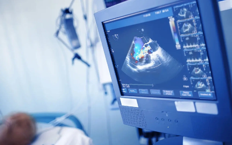
Transesophageal Echocardiography (TEE) is one of the advanced and accurate heart imaging methods that provides more comprehensive information than conventional echocardiography (from the chest) due to the removal of obstacles such as ribs or fat tissues. In this method, the transducer of the echocardiography device enters the esophagus through the mouth. Because the esophagus is located close to the heart and there are no obstacles such as ribs, skin or fatty tissues, it allows for very high resolution imaging and recording of detailed details of the internal structures of the heart. Easy accessShare content
Instagram Facebook X-twitter TelegramThe most important reasons for performing echocardiography
Doctors use transesophageal echocardiography for sensitive surgeries and important treatment decisions, especially when other methods do not provide enough information. Echo-Echo is a key tool in the management of heart disease and helps to maintain the patient's health in the best possible way.
- Providing very high resolution images due to the proximity of the transducer to the heart
- Cardiac evaluation before major surgeries or immediately after open heart surgery
- Ensuring the proper functioning of the heart and valves
- Exact examination of heart valve problems such as stenosis or insufficiency of aortic and mitral valves
- Important role in identifying blood clots inside the heart (especially in patients with atrial fibrillation or after stroke)
- providing sufficient and more accurate information about the structure or function of the heart than other imaging methods
- Diagnosis of structural abnormalities of the heart such as inter-atrial or ventricular holes
- Possibility of accurate diagnosis of complex diseases and appropriate treatment
- Effective in checking the performance of artificial valves
- Effective role in identifying abscesses or infectious masses in patients suspected of endocarditis (heart valve infection)
In what cases is echocardiography performed?
Echo of the heart or transesophageal echocardiography (TEE) is used when echocardiography from the chest cannot provide clear and sufficient images.
Know the types of esophageal echocardiography...
Each type of echo esophagus has its own use and benefits, and the cardiologist chooses the right type according to your needs. Familiarity with these types can help to better understand the applications of this advanced method.
2D transesophageal echocardiography (2D TEE)
This method is the most common type of echocardiography that provides two-dimensional images of the structure of the heart. Doctors use these images to evaluate the valves, cavities, and great vessels connected to the heart.
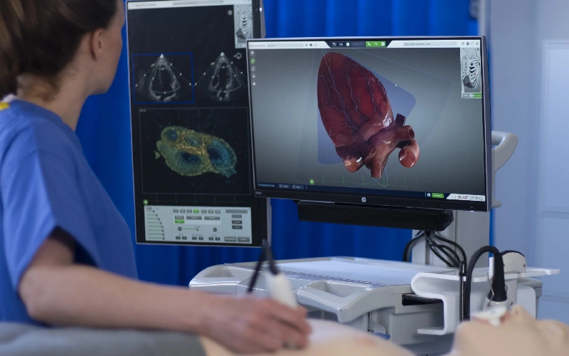
3D transesophageal echocardiography (3D TEE)
This type of echo provides three-dimensional and very detailed images of the heart, which are very useful in detecting small details such as valvular defects or heart wall defects. This type of echo is used in complex surgeries or to check the function of artificial valves.
Echo heart through Doppler (Doppler TEE)
In this method, the blood flow in the heart and main vessels is checked. This type of echo especially plays an important role in diagnosing insufficiency or stenosis of valves and hemodynamic disorders.
Stress TEE
In certain circumstances, echocardiography is performed under the influence of stress (pharmacological or physical) in order to check the heart's function during more activity. This method is used to evaluate coronary artery disease or evaluation before important surgeries.
Contrast TEE
By injecting a contrast agent, more details of the structure of the heart and blood vessels are displayed. This method is very effective for identifying blood clots or complex heart abnormalities.
Comparison of echocardiography through the esophagus with echocardiography through the chest
feature
Echo through the chest
Echo through the esophagusImage clarity
averageVery high
Required equipment
simplemore advanced
Need special preparation
does not haveCompulsory fasting
specialized applications limitedwider and more specialized
Familiarity with the steps and how to perform echocardiography through the esophagus
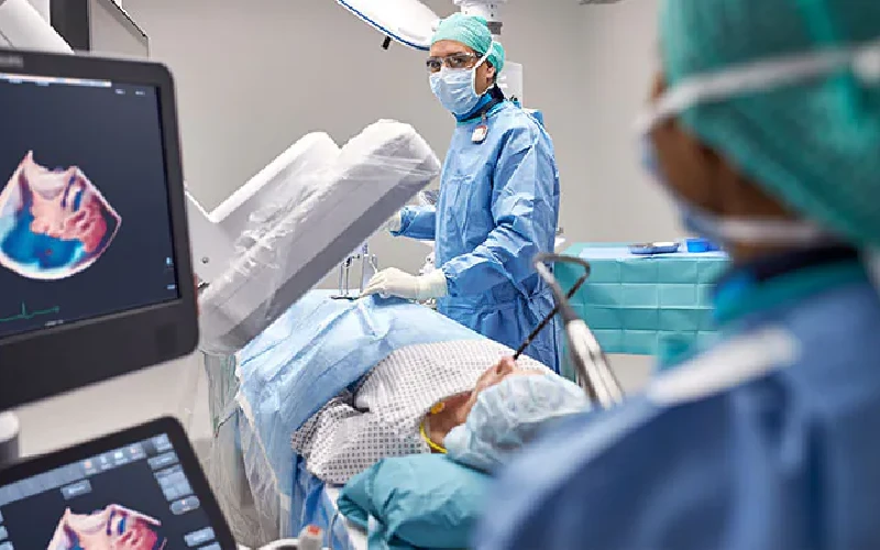
In this method, the transducer of the echocardiography device reaches near the heart through the mouth and esophagus to record clearer images of the inner parts of the heart. Doing this method includes several main steps:
- First, the patient should fast for at least 6 hours before the echo to reduce the risk of vomiting and side effects. Before starting, the doctor prepares the throat with a local anesthetic spray so that the insertion of the transducer does not cause discomfort. In most cases, a sedative is injected to make the patient feel comfortable during the procedure.
- During the echo, the patient lies on his side and the transducer is slowly inserted into the esophagus through the mouth. The doctor places the transducer in the right position to capture detailed images of the heart and surrounding structures. These images are obtained by moving the transducer in different directions and changing its angle. The entire imaging process usually takes between 20 and 40 minutes.
- After the work is done, the converter is slowly removed. The patient may experience a mild sore throat that resolves within a few hours. It is usually recommended that the patient avoids eating and drinking until the effect of anesthesia is completely removed.
Investigation of possible complications caused by echocardiography through the esophagus
Transesophageal echocardiography (TEE) is generally a safe and low-risk procedure, but like any other medical procedure, it may carry risks and complications. One of the common side effects of this method is a sore throat or a feeling of discomfort in the throat after inserting the mask, which is usually temporary and resolves very quickly on the first day. In rare cases, especially if the patient has underlying problems, it may cause serious injuries:
- Appearance of reactions to sedatives or local anesthesia (nausea, dizziness or allergies)
- Esophageal rupture Internal bleeding
- Disturbance in breathing
Note that these complications occur more in patients with complex health conditions or when referring to inexperienced doctors.
To reduce these risks, it is important for the patient to share all their medical records and drug sensitivities with the doctor before performing this procedure. Also, performing this procedure in specialized centers and under the supervision of experienced doctors can maximize its safety. In general, the benefits of this method in the accurate diagnosis of heart diseases far outweigh its risks.
General conditions of the patient during echocardiogram test
During the test, the patient usually lies on the bed and carefully maintains their body position to place the transducer in the esophagus. During the time when the doctor or echo technician inserts the transducer through the mouth into the esophagus and places it in the right position, the patient must be fully cooperative and especially calm when moving the transducer in the esophagus. The patient may have an unpleasant sensation such as a temporary sore throat or a feeling of fullness in the throat, but these discomforts are usually temporary and minor. After the test, the patient usually needs a short rest to wear off the sedative effects and return to normal. The general condition of the patient should be under the supervision of a specialist doctor so that any side effects can be identified and treated in time.
How long does a transesophageal echocardiogram take?
The duration of the esophageal echocardiogram is usually between 20 and 40 minutes. However, the entire process from the patient's entry into the echo room to preparation and post-procedure relaxation may take about 1 to 2 hours.
- The initial stage takes about 10 to 15 minutes (initial preparation of the patient including checking the vital signs, injecting a sedative if needed and using an anesthetic spray to numb the throat)
- The second step, depending on the complexity of the patient's condition, takes between 20 and 30 minutes (inserting the echo transducer into the esophagus through the mouth and imaging the heart)
After the imaging is completed, the transducer is slowly removed and the patient is monitored for a short period of time to wear off the sedatives.
What is the recovery period after echo through the esophagus?
The recovery period after transesophageal echocardiography (TEE) is usually short and without serious complications. This method is generally minimally invasive and the patient can return to his normal activities after a few hours.
Overall, the recovery period of this procedure is very short and most patients can return home and resume their normal activities on the same day.People who have received a sedative should avoid driving and doing activities that require concentration for the next day. Also, on the day of the echo, it is better for the patient to be under the supervision of another person in order to receive help in case of an unexpected problem.
Getting to know 6 advantages of echocardiography compared to other methods
- The ability to diagnose complex diseases such as blood clots in the left atrium
- The ability to diagnose heart infections (endocarditis)
- Ability to detect structural abnormalities of the heart
- Providing the possibility of a detailed examination of the function of heart valves and valve prostheses
- Essential use in cases of heart surgery that require detailed information
- Providing reliable results in patients with excessive weight or lung problems

How much does Echo Mary cost?
The cost of echocardiography depends on various factors and may vary in different medical centers. Some of the most important factors that affect the cost of this test are:
- Type of treatment center: Doing an echocardiogram in public hospitals is usually less expensive than in private centers. Specialized heart centers may also have different costs.
- Type of insurance: Many basic and supplementary insurances cover part of the cost of Echo Mary. The amount of insurance coverage depends on the type of contract and the limit of the insured's obligations.
- Equipment and technology used: Centers that use more advanced and newer devices may charge more.
- Receive ancillary services: If other tests or services such as sedation are provided along with echocardiography, the final cost will increase.
Factors that are considered in the interpretation of echocardiogram results
Interpretation of echocardiogram results can provide important information about your cardiovascular health. In an echocardiogram, images of the heart and valves are taken so that the doctor can accurately assess the heart's function, blood flow, and vascular condition.
In interpreting the result of echocardiography, the first thing that is checked is the size and shape of the heart. Changes in the size of the heart chambers can be a sign of problems such as heart failure or abnormal heart enlargement. Also, the condition and function of heart valves are evaluated. Valvular diseases, such as stenosis or insufficiency of the valves, which lead to impaired blood flow, are usually detected in this test.
Another important aspect of echocardiographic interpretation is the examination of heart muscle function. If the heart muscle is weak or does not contract properly, it may be a sign of conditions such as previous heart attacks or heart failure. Also, the blood flow in large veins and arteries is also checked. Impaired blood flow or the presence of blood clots can lead to serious problems such as heart attack or stroke. Finally, the images obtained from the echo esophagus can help identify congenital heart defects, such as a hole between the two chambers of the heart.
How long does it take for transesophageal echocardiography results to be ready?
For emergencies, such as detecting blood clots or checking for a heart infection, results are usually available immediately. In non-emergency cases, an official report including a complete analysis and treatment recommendations will usually be ready within 24 to 48 hours.
In most cases, the initial results can be interpreted immediately after the echo is performed by a specialist doctor. During an echocardiogram, the doctor views the images live and may provide you with basic information right away.
Although the initial analysis is quick, in some cases the recorded images may require a more detailed review by a cardiologist or radiologist. This process can take several hours to several days, especially if the images are referred to other doctors for more detailed review.
Edit Clone Comments read more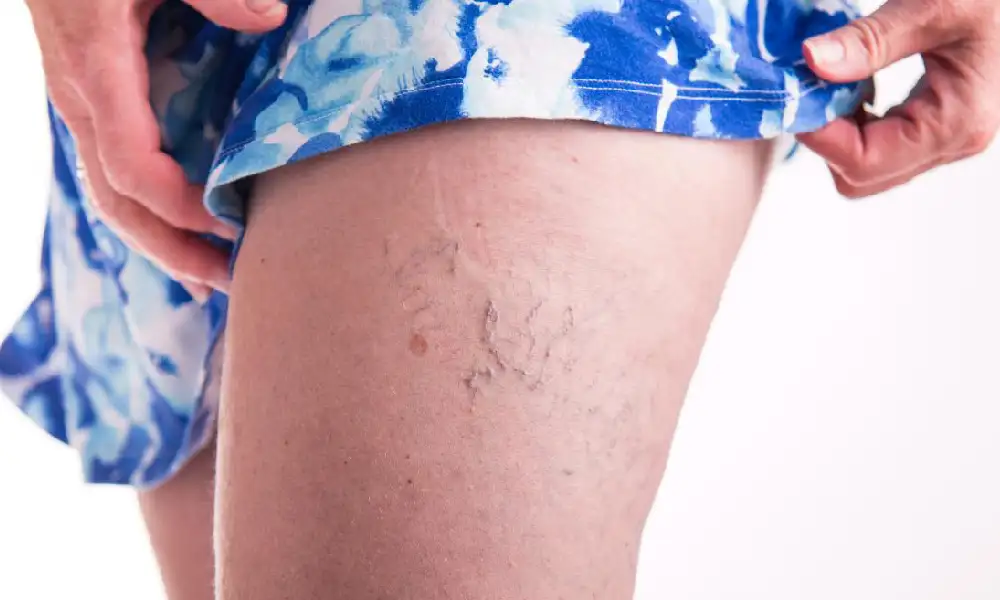 Why care about leg veins? Solutions to prevent vascular problems
02
April
Why care about leg veins? Solutions to prevent vascular problems
02
April
 10 effective ways to maintain heart health and prevent cardiovascular diseases
02
April
10 effective ways to maintain heart health and prevent cardiovascular diseases
02
April
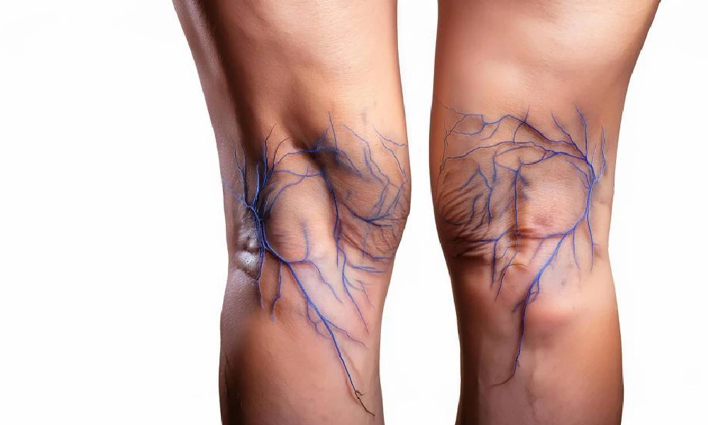 introduction of several home treatment methods for varicose veins
07
D
introduction of several home treatment methods for varicose veins
07
D
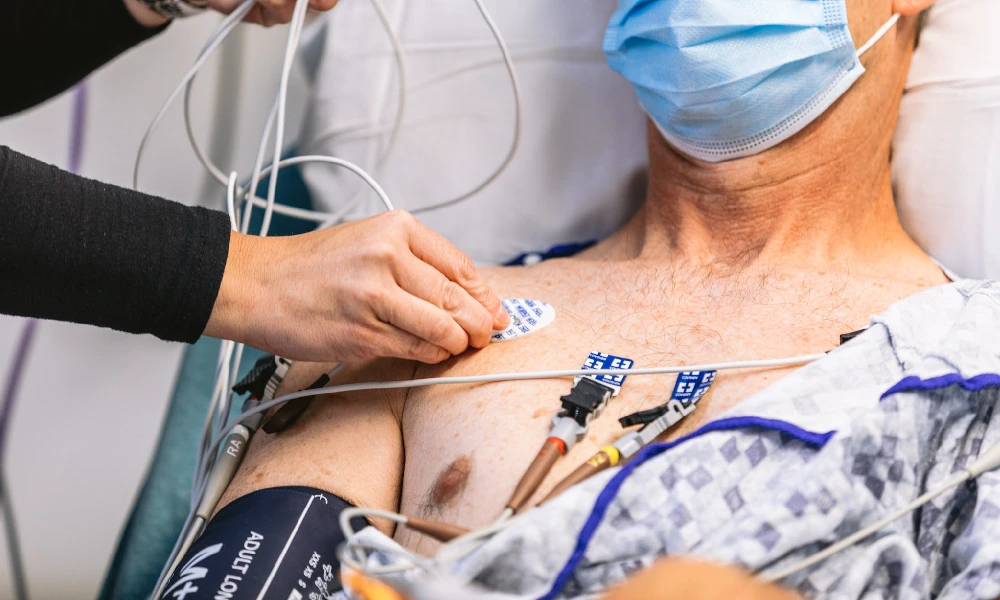 Preparation before taking an ECG; Should we be fasting to take an ECG?
07
D
Preparation before taking an ECG; Should we be fasting to take an ECG?
07
D
 The least dangerous methods of treating coronary artery occlusion
07
D
The least dangerous methods of treating coronary artery occlusion
07
D
 3-day apple weight loss diet
07
D
Your comments
0
0
votes
Article Rating
Subscribe to
Login
0 Comments
the oldest
the most recent
The most votes
Feedback (Feedback) inline
View all comments
3-day apple weight loss diet
07
D
Your comments
0
0
votes
Article Rating
Subscribe to
Login
0 Comments
the oldest
the most recent
The most votes
Feedback (Feedback) inline
View all comments
مقالات دیگر از Dr. Seyed Alireza Najafi

Review of 7 ways to prevent high blood pressure - Dr. Seyed Alireza Najafi
Review of 7 ways to prevent high blood pressure High blood pressure means that your heart is pumping blood around the body more forcefully. This extra pressure can damage blood vessels an...

What is normal blood pressure? - Dr. Seyed Alireza Najafi
What is normal blood pressure? Blood pressure is the force with which the blood presses against the walls of the vessels. This force is like water being forced out of a hose. The higher t...

How are pulmonary function tests? - Dr. Seyed Alireza Najafi
What are pulmonary function tests? Pulmonary function tests are a series of tests that help doctors evaluate how well your lungs are working. These tests can help diagnose and track lung...

Varicose vein treatment methods - Dr. Seyed Alireza Najafi
Types of varicose vein treatment methods Varicose veins are swollen and twisted veins that are often seen in the legs. These veins are created due to failure of venous valves and accumula...

Care after angiography at home Dr. Seyed Alireza Najafi
Care after angiography at home Dos and don'ts Care after angiography at home is very important for the healing process to go well and to avoid possible complications. By following the ti...

Treatment of hand pain after angiography Dr. Seyed Alireza Najafi
Know the techniques for treating hand pain after angiography! Since today angiography is considered one of the most important and reliable methods in the field of diagnosis and treatment...

cause of chest pain; From heart diseases to lung and digestive problems
What is the cause of chest pain? Introduction of prevention and treatment methods The onset and duration of any disease in the body is associated with the development of symptoms and com...

treatment of varicose veins with laser; Say goodbye to varicose veins
Getting to know the methods of laser treatment of varicose veins Varicose veins, especially varicose veins of the legs, are among the most common diseases that, in addition to creating a...

7-day hydrotherapy regimen; Is it worth losing 3 to 4 kilos?
Seven-day hydrotherapy diet, the best fast weight loss diet The effect of any type of diet on a person depends on his physical condition, lifestyle and metabolic characteristics of his bo...

Fatty liver treatment diet; 3 of the best fatty liver diets
Is it possible to treat fatty liver with diet? Fatty liver treatment diet Today, fatty liver is one of the common diseases. One of the questions that most people with this disease have is...

How to take an ECG | The easiest way to diagnose heart diseases
How to take an ECG? Taking an electrocardiogram is a simple, painless and risk-free test that a doctor prescribes to check the electrical activity of the heart. The results of an electro...

Women's heart echo The importance of women's echo and its differences with men's heart echo
What is the difference between women's heart echo and men's echo? Nowadays, women may need an echocardiogram for several reasons, including congenital heart diseases, pregnancy complicati...

SEO site management services
Search engine optimization (SEO) services help you to rank your website higher in the search results of Google and other search engines.

Implementation of branding and digital advertising
Digital branding means creating a strong and distinctive brand in the digital space for a company or a specific product. This process involves using digital methods and strategies to build and strengthen the brand.

Website design for companies and businesses
Website design for companies and businesses can help you have a stronger online presence and attract more customers. Contact us for more information.